4–9 Month Timeline for Dental Bone Grafts in Canada: What to Expect


A bone graft for implants rebuilds jaw bone that’s too thin, too short, or too soft to hold an implant securely, using it as a scaffold your body fills in with new bone over several months. Most patients need one when a tooth has been missing for a while or was lost to gum disease. Healing typically runs 4 to 9 months depending on the graft type, and success rates are generally strong when risk factors like smoking are managed. We walk through the details below.
TL;DR:
Most grafts require four to six months to consolidate, with larger or sinus lift procedures often needing six to nine months before implants can be placed.
Smoking and uncontrolled health conditions significantly reduce graft success rates, so cessation during healing is highly recommended.
The choice of graft material—autograft, allograft, xenograft, or alloplast—depends on defect size, location, and surgeon experience, affecting healing time and costs.
Grafted sites tend to behave like natural bone long-term, but ongoing hygiene and regular checkups are essential to maintain implant stability.
A CBCT scan is critical for accurate assessment and planning, and treatment costs vary based on material, procedure complexity, and whether staged or simultaneous approaches are used.
Table of Contents
What Is a Dental Bone Graft, and Why Does It Matter for Implants?
An implant needs a certain amount of bone width and height to stay stable for decades, not just months. When that bone is missing, a graft fills the gap with material your body recognizes as a scaffold. Over time, your own bone cells grow into it, gradually replacing the graft with living, vascularized bone strong enough to anchor a titanium implant.
Why does bone go missing in the first place? Once a tooth is extracted, the jawbone that used to support it starts to resorb almost immediately, because bone needs the mechanical stimulation of a tooth root to maintain its shape. Wait a year or two after an extraction without addressing the socket, and you’ll often lose meaningful width and height in that ridge. Gum disease works the same way over a longer timeline, slowly eating away at the bone that surrounds your teeth. Trauma, long-term denture wear, and developmental factors can produce the same result.
Before recommending a graft, your dentist needs to know exactly how much bone you have and where. That’s where cone beam computed tomography (CBCT) comes in. Unlike a flat two-dimensional X-ray, CBCT imaging builds a 3D map of your jaw, letting clinicians measure ridge width and height down to the millimeter and plan the exact graft site and implant angle before any incision is made.
Common scenarios where grafting comes up during implant planning include:
A tooth extracted months or years ago without socket preservation, leaving the ridge narrow or collapsed.
Advanced gum disease that has eaten away supporting bone around neighboring teeth.
Trauma or infection that destroyed bone at the site.
Long-term denture use, which accelerates bone loss because there’s no tooth root stimulating the ridge.
A ridge that’s simply too thin or too short on a CBCT scan to achieve primary implant stability, even without an obvious cause.
If you’ve already lost bone and are wondering whether implants are still on the table, they usually are. Bone loss doesn’t automatically rule out implants; it just means grafting typically becomes part of the plan.
What Are the Different Types of Bone Grafts?
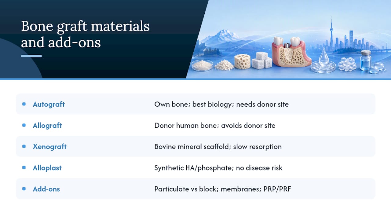
Your options fall into four material categories, plus a few structural and biological add-ons that change how the graft behaves. A clinical review of grafting materials covers all four in detail, and no single material wins outright. The right choice depends on the size of your defect, where it’s located, and what your surgeon has the most experience using.
Autograft means bone taken from your own body, usually the chin, the back of the jaw, or occasionally the hip. It has the best biological potential because it carries live cells and growth factors, but it requires a second surgical site, which means extra healing and some discomfort at the donor location.
Allograft is processed human bone from a tissue bank, typically freeze-dried or demineralized before use. It avoids a second surgical site entirely and remains one of the most common choices for ridge preservation, largely because it integrates predictably without the added morbidity of harvesting your own bone.
Xenograft comes from animal sources, most often bovine bone that’s been processed to remove all cellular material, leaving behind a mineral scaffold. It resorbs slowly, which makes it useful when you want the graft to hold its shape and volume for an extended period, such as under a sinus membrane.
Alloplast refers to synthetic materials like hydroxyapatite or calcium phosphate ceramics. These are manufactured rather than harvested, which means no disease transmission risk and unlimited supply, though they sometimes integrate more slowly than biological options.
On top of material choice, your surgeon decides between particulate grafts (granules packed into a defect) and block grafts (a solid piece shaped and screwed into place). Particulate material is the standard approach for most defects because it’s easier to place, contours to irregular shapes, and generally recovers faster. Block grafts get reserved for severe vertical or horizontal defects, often larger than 5 millimeters, where a solid structural piece is needed to rebuild significant lost volume.
Membranes often get placed over the graft to keep it contained and to block soft tissue from invading the space before bone can form, a technique called guided bone regeneration. Resorbable membranes dissolve on their own; nonresorbable membranes need a small follow-up procedure for removal. Some clinicians also add platelet-rich plasma (PRP) or platelet-rich fibrin (PRF), concentrated from your own blood, to try to speed early healing.
All of these materials are approved and routinely used in Canadian dental and oral surgery practices, subject to standard Health Canada medical device regulations for processed tissue and synthetic products.
Pro Tip: Ask your provider which specific material they’re using and why for your defect. “Bone graft” isn’t one product. It’s a category, and the material matters for both healing time and cost.
What Happens During a Bone Graft Procedure?
The approach depends on timing relative to any extraction, and there are three main pathways.
Socket preservation happens immediately after a tooth extraction, placing graft material directly into the empty socket before it has a chance to collapse. This is the least invasive option and often the most convenient, since it piggybacks on a procedure you’re already having.
Staged augmentation is used when significant bone loss has already occurred and needs to be rebuilt before an implant can go in at all. The graft goes in on its own, heals for months, and the implant follows later as a separate procedure.
Simultaneous placement combines the implant and the graft in one surgery, typically when there’s enough existing bone for primary stability but a smaller defect around the implant needs filling.
A typical particulate graft appointment follows this sequence:
Local anesthesia numbs the site; some patients also opt for oral or IV sedation if anxious about the procedure.
The surgeon accesses the bone site, either through the fresh extraction socket or by making a small incision in healed tissue.
Graft material is packed into the defect and, in most cases, covered with a membrane.
Sutures close the site, and imaging or a clinical check confirms placement.
Block grafts add steps: the surgeon first harvests bone from a donor site (often the chin or the back of the lower jaw), shapes it to fit the defect, and secures it with small titanium fixation screws so it can’t shift during healing. Ridge expansion, used when the ridge is thin but has adequate height, involves gently spreading the bone with specialized instruments and packing graft material into the resulting space, sometimes allowing an implant the same day.
Postoperative discomfort is usually described as manageable, more akin to a tooth extraction than a major surgery, and typically peaks in the first 48 to 72 hours before tapering off. Swelling is common for 3 to 5 days. Most patients return to normal activity within a few days, though block graft donor sites can feel sore for a week or more.
CBCT scans anchor the entire process, both before surgery to plan the defect size and material volume, and afterward to confirm graft integration ahead of implant placement.
Sinus Lift vs. Bone Graft: What’s the Difference?
A sinus lift is a specific type of bone graft used only in the upper back jaw, where the sinus cavity sits directly above the tooth roots. Regular ridge grafting builds bone laterally or vertically along the jaw; a sinus lift gently raises the floor of the sinus membrane itself and packs graft material into the new space beneath it, creating height where the sinus has expanded downward over the years.
There are two surgical approaches:
Lateral window technique: the surgeon opens a small window in the side of the jawbone to access and lift the sinus membrane directly. This is used for larger height deficits and generally requires healing before implant placement, often 6 to 9 months or longer.
Crestal (osteotome) technique: a less invasive approach through the implant site itself, used when only a modest lift is needed. When residual bone height is adequate, this method sometimes allows the implant to go in during the same appointment as the lift.
The choice between the two comes down to how much height you’re missing. A few millimeters might mean a crestal lift and simultaneous implant placement. A larger deficit typically means the lateral window approach and a staged healing period before the implant stage begins.
It’s also worth knowing these aren’t mutually exclusive. Some patients need both a sinus lift for vertical height and separate ridge grafting for horizontal width, particularly if they’ve been missing teeth in that region for years. Your surgeon will map this out on your CBCT scan rather than guessing during the procedure itself.
How Long Does Bone Graft Healing Take Before Implant Placement?
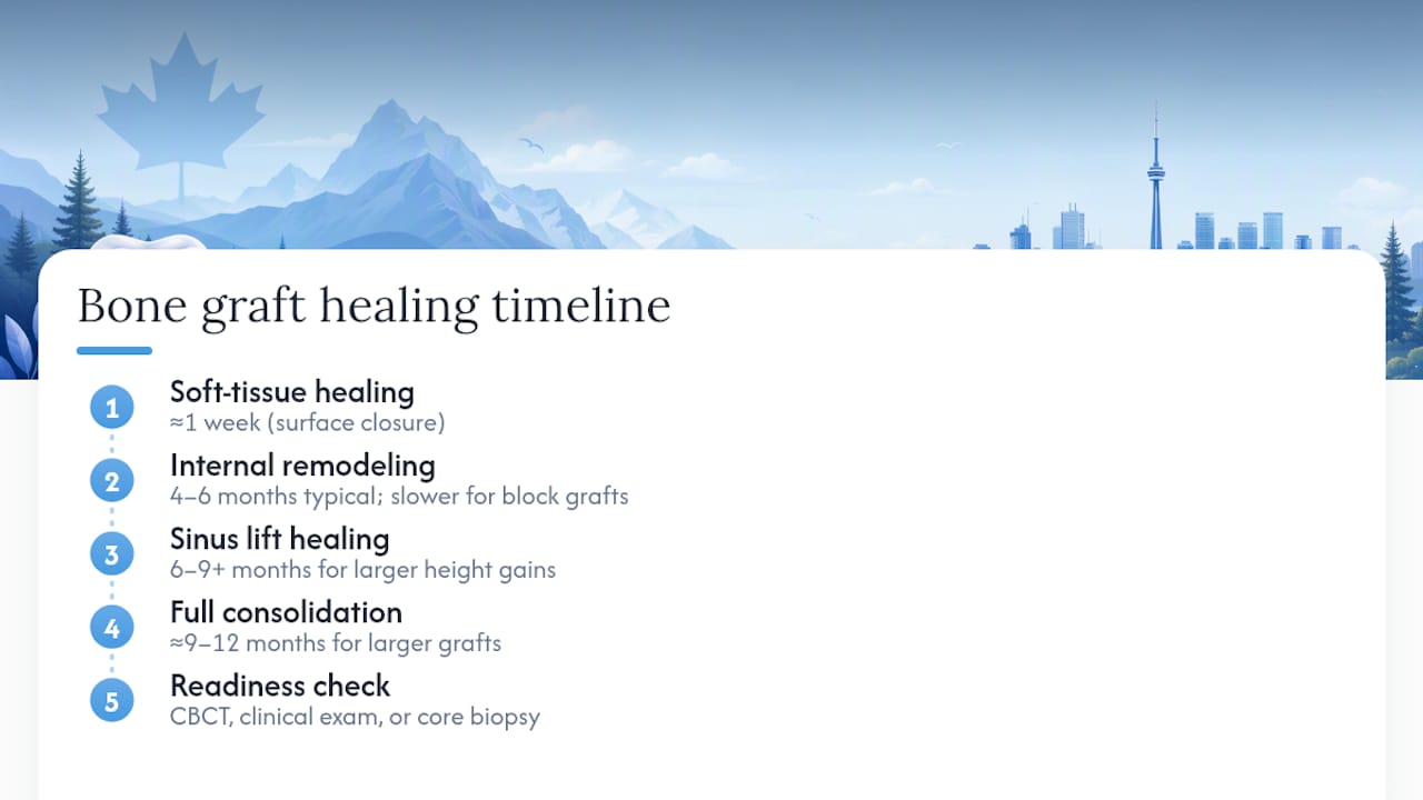
Healing time depends heavily on graft type and size, and the research on this is more specific than most patients expect. Particulate grafts used for socket preservation or moderate ridge defects generally need 4 to 6 months to consolidate enough for implant placement. Block grafts, because they involve a larger volume of material and a slower vascularization process, often need 6 months or more. Sinus lifts using the lateral window approach frequently run 6 to 9 months or longer, particularly with larger height gains.
A randomized controlled trial comparing healing intervals after socket preservation with demineralized freeze-dried bone allograft found nearly 50% more new vital bone at the longer healing point: 47.41% new bone at 18 to 20 weeks compared with 32.63% at 8 to 10 weeks.
That trial matters because it gives an actual histologic answer to a question patients ask constantly: does waiting longer really make a difference, or is it just clinical caution? The data says waiting genuinely produces more mature, viable bone, not just a longer calendar. It’s a big part of why many surgeons resist rushing implant placement even when a graft site looks healed on the surface.
Surface appearance can be deceiving. A graft site can look fully healed in the mouth while the bone underneath is still remodeling internally, which is why clinicians rely on more than a visual check. Readiness is typically confirmed through a combination of a follow-up CBCT scan measuring density and volume, a clinical exam checking that the tissue is firm and stable, and occasionally, in ambiguous cases, a small core biopsy taken at the time of implant placement to directly assess bone quality.
Initial soft tissue healing after a graft is fast, often within about a week, but that’s not the same as the graft being ready to support an implant. Full consolidation, the point where the grafted material has been substantially replaced by your own living bone, can take several months up to 9 to 12 months for larger grafts. If your provider tells you to wait longer than you expected, it’s often because the evidence supports that extra patience translating into a more durable result, not because of unnecessary delay. For a broader view of how grafting fits into the overall implant sequence, the typical implant timeline runs anywhere from 3 to 12 months from first consult to final crown, largely driven by whether grafting is needed and what kind.
What Are the Risks and Success Rates for Bone Grafts?
Bone grafting is a well-established procedure, but it’s still surgery, and it carries a specific set of risks worth understanding before you commit.
The most common complications include:
Infection at the graft site, which can compromise integration if not caught early.
Graft exposure, where the membrane or graft material becomes visible through the gum tissue, usually from a small tear or incomplete closure.
Donor-site morbidity after autograft harvesting, meaning pain, swelling, or occasionally altered sensation at the chin or jaw donor location.
Sinus membrane perforation during sinus lift procedures, which is usually manageable during surgery but can occasionally delay the lift.
Partial or complete graft failure, where the material doesn’t integrate as planned and needs to be redone.
Certain patient factors substantially change these odds. Smoking is one of the most consistently identified risk factors for reduced graft take and lower implant success, largely because nicotine restricts blood flow to healing tissue right when new blood vessel growth matters most. Poorly controlled diabetes, a history of head and neck radiation, and active untreated periodontal disease all raise complication risk as well. If you smoke, understanding how it affects implants specifically before your surgery date is worth the read.
Reported success and graft “take” rates in the literature run high overall, though exact figures vary by study design, graft type, and defect size, which makes a single universal number misleading. Particulate grafts for straightforward ridge preservation tend to have the most predictable outcomes; larger block grafts and sinus lifts carry somewhat more variability simply because they’re addressing bigger deficits.
When a complication does occur, treatment usually starts conservatively: antibiotics for infection, minor tissue adjustment for exposure, or extended healing time if integration is slower than expected. Outright graft failure isn’t common, but when it happens, re-grafting is generally straightforward and doesn’t preclude a successful implant down the road.
Pro Tip: If you smoke, ask your provider about a cessation plan before your graft date, not after. Even a temporary pause around the surgical window can meaningfully improve your healing odds.
How Much Does a Bone Graft for Implants Cost?
Bone graft pricing varies more than most other dental procedures because so many variables feed into the final number. The graft material itself is one factor: allograft and synthetic options are typically less expensive than block autografts, which require a second surgical site and more surgical time. Whether the procedure is done by a general dentist or an oral surgeon can shift cost, as can whether sedation beyond local anesthesia is used.
Other major cost drivers include:
Graft type and volume: particulate socket grafts are generally less costly than block grafts or sinus lifts, which involve more material and surgical complexity.
Simultaneous vs. staged treatment: combining a graft with implant placement in one visit can sometimes reduce total fees compared with two separate surgical appointments.
Imaging requirements: a CBCT scan for planning is typically billed separately and is a necessary part of an accurate quote, not an optional add-on.
Sedation choice: local anesthesia alone costs less than IV or oral sedation for anxious patients or more extensive procedures.
Because of this range of variables, providers typically quote grafting as a price band rather than a flat fee, and any number you see online without a CBCT-based assessment of your specific case should be treated as a rough estimate at best.
Insurance coverage for bone grafting varies by plan, and many Canadian dental insurance policies cover at least a portion of grafting when it’s medically necessary for implant placement, though coverage details differ significantly between providers. If you’re eligible for the Canadian Dental Care Plan (CDCP) or the Interim Federal Health Program (IFHP), it’s worth confirming what’s covered before your consult so there are no surprises at billing. Clinics that offer direct insurance billing simplify this considerably, since you’re not stuck paying the full amount up front and waiting on reimbursement.
The most reliable way to get an accurate number is a CBCT-based consult where your provider can actually see your bone volume and defect size, rather than guessing off a general price list.
How to Prepare for a Bone Graft and What to Ask Your Provider
Preparation starts weeks before your surgery date, not the morning of.
If you smoke, stopping or at least significantly cutting back in the weeks surrounding your graft date measurably improves healing odds. Review all current medications with your provider, since certain blood thinners and some osteoporosis medications can affect bone healing or bleeding risk and may need adjustment. Arrange transportation and someone to help you home if you’re opting for sedation, since driving yourself isn’t safe afterward.
Aftercare in the days following surgery generally includes:
A soft diet for the first several days, avoiding anything crunchy, hot, or that requires significant chewing near the graft site.
Modified oral hygiene, often gentle rinsing rather than brushing directly over the surgical area for the first week.
A pain control plan, usually over-the-counter anti-inflammatories, sometimes supplemented with a short prescription course for larger grafts.
Follow-up imaging at intervals your provider sets, typically checking progress at a few months in and again closer to the anticipated implant date.
Before you commit to a treatment plan, bring these questions to your consult: Who will actually perform the surgery, a general dentist or an oral surgeon? What specific graft material is being used, and why that one for your defect? What’s the realistic timeline from graft to implant to final restoration? What happens if the graft doesn’t take as expected, and is there a contingency plan? What’s the full cost estimate, including imaging, and how does insurance or CDCP/IFHP factor in? A rinse routine that respects the healing graft site also matters more than people expect; proper technique and timing for mouthwash after extraction applies just as directly to graft sites.
Pro Tip: Write your questions down before the consult. It’s easy to forget half of them once you’re sitting in the chair, and the answers matter for both your timeline and your budget.
Contact your provider promptly if you notice increasing pain after the third day, visible pus or unusual swelling, a fever, or if the graft material or membrane appears to be coming through the gum tissue. None of these are common, but catching them early makes treatment far simpler.
What Are the Long-Term Outcomes After a Bone Graft?
Once a graft has consolidated and an implant is successfully placed on top of it, long-term outcomes are generally favorable, and the grafted bone tends to behave like the surrounding native bone from that point forward. The implant itself now provides the mechanical stimulation that keeps that bone maintained, similar to how a natural tooth root does.
That said, grafted sites aren’t entirely maintenance-free. Regular dental checkups let your provider monitor bone levels around the implant over time, typically through periodic imaging every few years rather than annually once things are stable. Gum health around the implant deserves particular attention, since peri-implant disease (inflammation around an implant, similar in concept to gum disease around a natural tooth) can slowly erode the very bone the graft worked to rebuild.
Daily habits matter more here than most patients expect. Consistent flossing or interdental brushing around the implant crown, avoiding tobacco long-term, and keeping any underlying conditions like diabetes well controlled all support the durability of that grafted bone for years to come. Patients who maintain good hygiene and attend regular checkups generally see grafted implant sites remain stable well beyond the initial healing period, often for decades. The bone that seemed like the hardest part of the whole process tends to become, over time, simply part of a healthy jaw.
Are There Alternatives to Bone Grafting for Implant Sites?
Not every patient with insufficient bone needs a traditional graft, and a few adjunctive techniques can sometimes shorten the path to an implant.
Ridge expansion is one option when the ridge is thin but adequately tall. Rather than adding bulk bone material and waiting months, the surgeon uses specialized instruments to gently widen the existing ridge, then places graft material into the newly created space, sometimes allowing implant placement in the same visit. This works because the existing bone is being repositioned and expanded rather than entirely rebuilt from scratch, which can save months compared with staged grafting when the anatomy cooperates.
Distraction osteogenesis is a more involved technique used for larger vertical deficiencies. A small device is surgically placed to gradually separate a segment of bone, and the body naturally fills that widening gap with new bone over several weeks, essentially growing bone in place rather than importing material. It’s a more specialized approach, typically reserved for significant defects where traditional grafting would need an unusually large volume of material.
Short or narrow-diameter implants sidestep the bone deficiency question entirely in select cases, using implant designs that fit the bone you already have rather than building more bone to fit a standard implant. This isn’t right for every situation and depends heavily on bite forces and location, but it’s worth discussing if you’re hoping to avoid grafting altogether.
None of these replace grafting universally. They expand the toolkit for specific anatomical situations, which is exactly why a thorough CBCT-based assessment matters before assuming a graft is your only path forward.
Bone grafting is approached as a planning process, often starting with a CBCT scan to assess bone volume and plan treatment. Discussions about graft material, timelines, and costs typically occur before scheduling any procedures. New patients are accepted, and direct insurance billing plus participation in government dental programs may be available. Same-day treatment and emergency care options can provide quicker access to care when needed.
Ready to Plan Your Implant and Graft Consultation?
A CBCT-based consult can provide information about bone volume, possible graft needs, and a cost estimate before committing to treatment. Unlike piecing together information from articles and general price lists, you leave the appointment with a plan specific to your jaw, not a national average.
The clinic accepts new patients, offers direct insurance billing, and participates with government dental programs. Same-day and emergency appointments may also be available for urgent dental concerns.
Here’s what to do next:
Book a CBCT consult through our implants page to get precise imaging of your jaw bone.
Check your insurance eligibility, including CDCP or IFHP coverage, before your appointment so pricing questions are answered up front.
Request an itemized estimate covering graft material, imaging, and the implant itself, so there are no surprises later.
Visit our full services page to see how grafting and implants fit alongside the rest of your dental care, or reach out directly to get a consult on the calendar.
Key Studies and Clinical Resources
The healing timeline claims in this article are grounded in a randomized controlled trial on healing time and new bone formation, which remains one of the clearest histologic looks at why longer consolidation windows produce more mature bone. For a broader view of graft material choices, the overview of autografts, allografts, and synthetic materials covers the clinical tradeoffs in more depth than any single blog post can.
For patient-facing explanations in plain language, the Cleveland Clinic’s guide to dental bone grafts is a solid reference, and the review of CBCT applications in implant dentistry explains why 3D imaging has become the standard for grafting and implant planning.
This article is general information, not a substitute for advice from a qualified doctor. Consult a qualified healthcare professional about your own circumstances before acting on anything here.
Sources
FAQ
How Painful Is Bone Grafting for Dental Implants?
Most patients describe the discomfort as comparable to a tooth extraction, manageable with over-the-counter pain relievers and typically peaking in the first 48 to 72 hours. Block grafts involving a donor site tend to be somewhat more uncomfortable than particulate grafts.
How Long Do Bone Grafts Last for Dental Implants?
Once fully consolidated and topped with a successful implant, grafted bone behaves like your natural bone and can last for decades with good oral hygiene and regular checkups. It isn’t a temporary fix; it becomes a permanent part of your jaw structure.
How Much Does an Implant Bone Graft Cost?
Cost varies widely based on graft material, size, and whether sedation or a separate donor site is involved, which is why providers typically quote a range rather than a single figure after reviewing your CBCT scan. Mersaldental offers direct insurance billing and accepts CDCP and IFHP, and current estimate details are available by booking a consult through our implants page.
How Successful Is Bone Grafting for Implants?
Success rates reported in the clinical literature run high overall for particulate grafts used in routine ridge preservation, with somewhat more variability for larger block grafts and sinus lifts due to bigger defect sizes. Smoking, uncontrolled diabetes, and active gum disease are the factors most consistently linked to lower success.
How Long Does It Take for a Bone Graft to Heal Before an Implant?
Particulate grafts generally need 4 to 6 months, block grafts often need 6 months or more, and sinus lifts using the lateral window technique frequently run 6 to 9 months or longer. Your provider confirms readiness with a follow-up CBCT scan and a clinical exam rather than a fixed calendar date alone.
Recommended
Comments