1–8 µSv to 1,073 µSv: Dental X Ray Safety in Canada Explained

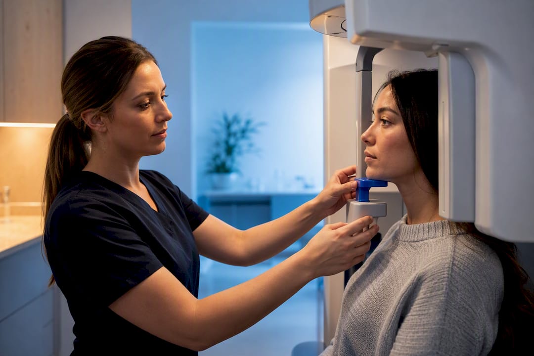
Dental X-rays are safe when they’re clinically justified and taken with dose-optimization practices like digital sensors and rectangular collimation, the standard known as ALARA (As Low As Reasonably Achievable) guided by Health Canada’s Safety Code 30. A single intraoral X-ray delivers roughly 1 to 8 microsieverts (µSv) of radiation, less than a day’s worth of natural background exposure most people absorb just living on Earth.
TL;DR:
Digital sensors and rectangular collimation significantly reduce dental X-ray doses, often to less than a day’s natural background exposure for intraoral images.
Evidence suggests that the risk of harm from routine dental X-rays is very small, with studies limited by biases and outdated technology that delivered higher doses.
Dentists are guided by Safety Code 30 and ALARA principles, emphasizing justification for each X-ray and minimizing dose through equipment, technique, and shielding, especially for children.
Patients should ask about necessity, lower-dose options, and safety practices, as dose reduction techniques like collimation and proper justification provide more safety than shielding alone.
Table of Contents
Understanding Dental X-Ray Safety and Radiation Doses by Image Type
We know patients often picture all dental X-rays as the same blast of radiation. They’re not. The dose depends entirely on which image your dentist takes, and the differences matter more than most people realize.
Intraoral X-rays are the bite-wing and full-mouth periapical images taken with a small sensor placed inside your mouth. These are the most common dental X-rays and also the lowest dose, typically 1 to 8 µSv per image.
Panoramic X-rays capture your entire jaw, teeth, and sinuses in one sweeping image from outside the mouth. Doses run higher than intraoral shots, generally landing around 4 to 30 µSv depending on the machine and settings.
Cephalometric X-rays, used mainly for orthodontic planning, capture a side profile of the skull and jaw. These come in lower, typically around 2 to 3 µSv, since the beam covers a wider area but at lower intensity.
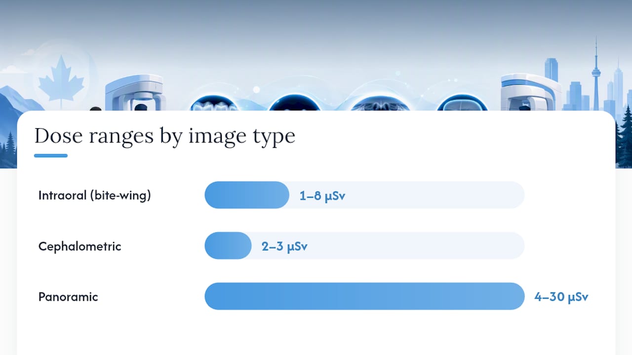
Cone-beam computed tomography (CBCT) is the outlier. It produces a 3D image used for implant planning, complex root canals, or surgical assessment, and the dose range is wide: 11 to 1,073 µSv, depending on the field of view, scan volume, and settings the dentist chooses. A small, targeted CBCT scan of a single tooth sits at the low end. A full-jaw scan with a large field of view sits at the high end, which is exactly why dentists are trained to select the smallest field of view that still answers the clinical question.
Here’s how those numbers stack up against a familiar benchmark:
Imaging type | Typical effective dose | Comparable to |
Intraoral (bite-wing) | 1–8 µSv | A few hours of natural background radiation |
Panoramic | 4–30 µSv | Less than one day of background radiation |
Cephalometric | 2–3 µSv | A few hours of background radiation |
CBCT (small field) | 11 µSv | A few days of background radiation |
CBCT (large field) | Up to 1,073 µSv | Roughly one to a few months of background radiation |
Why does “effective dose” matter as a concept rather than just a number? It’s the metric radiologists and health agencies use to compare very different imaging techniques on a common scale. The IAEA frames effective dose as a comparison tool, not a personal risk calculator. It lets a dentist weigh a panoramic image against an intraoral series, or compare dental imaging against a chest X-ray, using the same unit of measurement. That’s useful for planning care. It’s less useful for predicting what will happen to any one patient, a distinction that matters a great deal in the next section.
What the Evidence Says About Dental X-Ray Risks
The honest answer is that the evidence for real harm from dental X-rays at typical doses is thin, and what exists needs careful reading rather than alarm. A peer-reviewed review published in PMC looked at health effects tied to dental diagnostic X-ray exposure and found a handful of studies suggesting small associations with meningioma and thyroid cancer. The review’s own authors flagged significant limitations in that body of research, including recall bias (patients trying to remember X-ray history from years or decades earlier) and confounding factors that make cause and effect hard to isolate.
This is where the linear-no-threshold (LNT) model comes in. It’s the working assumption regulators use: that any radiation dose, no matter how small, carries some theoretical risk that scales down proportionally as the dose decreases. The model works reasonably well at the moderate-to-high doses seen in radiation therapy or major occupational exposure. At the microsievert doses typical of dental imaging, the model’s predictions become far shakier, because population-level statistical models struggle to detect an effect that small against the noise of everyday cancer risk from all other causes combined.
Statistic callout: A single intraoral dental X-ray (1 to 8 µSv) delivers a small fraction of the dose from one chest X-ray, and less radiation than most people absorb from natural background sources over the course of an ordinary day.
Put another way, the dose from routine dental imaging sits so far down the exposure scale that translating it into an individual cancer risk number is scientifically dubious. Health agencies use effective dose figures to compare and optimize techniques, not to tell a specific patient their personal odds of developing disease from one bite-wing series. That distinction gets lost in a lot of consumer health writing, and it’s worth restating plainly: population models are built for populations, not for predicting your outcome from one visit.
A few things worth holding onto from the research:
The associations reported in the PMC review were small and inconsistent across studies, not a confirmed causal link.
Recall bias is a serious problem in this research area, since most studies rely on patients self-reporting X-ray history from years earlier.
Modern digital sensors deliver up to 80% less radiation than older film-based systems, which means even the historical studies may not reflect current dental practice.
Dose optimization (not avoidance of necessary imaging) is what actually reduces whatever small theoretical risk exists.
None of this means dental X-rays carry zero risk, and no credible source claims that. It means the risk at these dose levels is small enough that untreated dental disease, missed fractures, or undiagnosed infections are almost always the larger threat to your health. That’s the trade-off dentists are trained to weigh every time they order an image.
How Safety Code 30 and ALARA Govern Dental Imaging
Canada regulates dental radiography through Safety Code 30, Health Canada’s national guidance document for the installation, use, and control of dental X-ray equipment. It’s not a suggestion. It’s the framework provincial regulators and dental colleges build their own rules around, covering everything from how equipment is calibrated to how staff are trained.
Two principles sit at the center of that framework: ALARA (As Low As Reasonably Achievable) and its close cousin ALADA (As Low As Diagnostically Acceptable). Both boil down to the same two-step logic every dentist is trained to apply before pressing the exposure button:
Justification. Is this X-ray actually necessary right now, based on your symptoms, history, or treatment plan? An image taken “just to check” without a clinical reason fails this test.
Optimization. Once an X-ray is justified, what’s the lowest dose that still produces a diagnostically useful image? This is where equipment settings, technique, and shielding decisions come in.
Safety Code 30 translates those principles into concrete facility obligations. Clinics operating X-ray equipment are expected to maintain:
A documented radiation protection program with clear staff responsibilities.
Regular equipment quality assurance and maintenance checks, including acceptance testing when new machines are installed.
Operator training specific to the equipment in use, updated as technology changes.
Guidance on thyroid shielding, applied when it doesn’t interfere with the diagnostic quality of the image, with particular attention for children.
Record-keeping practices that support both patient safety and regulatory accountability.
Pediatric patients get special weight in this framework for good reason. Children’s tissue is estimated to be roughly 32 times more radiosensitive than adult tissue, which is why justification and optimization steps aren’t just best practice for kids. They’re the difference between responsible imaging and unnecessary exposure at an age where the stakes are genuinely higher.
Dose-Reduction Techniques and Questions to Ask Your Dentist
Modern dental offices have several concrete tools to shrink radiation exposure without sacrificing diagnostic value, and the most effective ones aren’t the ones patients usually assume; for a broader perspective, see the role of X-rays in chiropractic care.
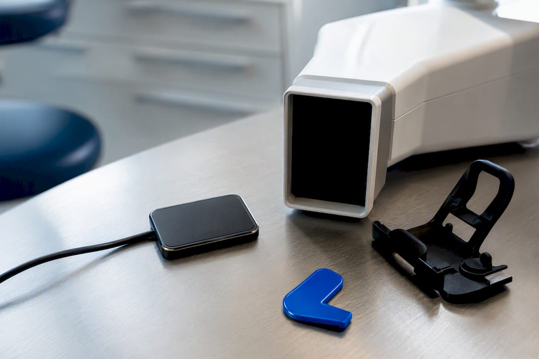
Digital sensors replace older film systems and cut required exposure time dramatically. Rectangular collimation, which shapes the X-ray beam to match the size of the sensor rather than blasting a wider circular field, reduces the exposed tissue area substantially. According to Safety Code 30 guidance, this single technique often does more to lower integral dose than patient shielding alone, since it stops excess radiation from ever reaching tissue outside the diagnostic target in the first place. Receptor holders keep the sensor stable and correctly positioned, which prevents the blurry or misaligned images that lead to retakes, and every retake is an avoidable second dose.
For panoramic and CBCT imaging, dentists optimize kVp (beam energy), mA (beam intensity), and exposure time for each patient’s size and clinical need rather than using one blanket setting for everyone. CBCT scans specifically should be limited to the smallest field of view that still captures the diagnostic information needed. A documented justification explaining why a lower-dose image wouldn’t answer the clinical question is considered good practice for every CBCT ordered.
Patients don’t need technical knowledge to advocate for themselves here. A few direct questions can confirm your dentist is applying these principles properly:
“Why is this X-ray needed today, specifically?”
“Is there a lower-dose imaging option that would still answer the question?”
“Is a CBCT scan actually necessary for my case, or would a standard image work?”
“What steps do you take to minimize exposure for children?”
“How often is your equipment checked or calibrated?”
Pro Tip: A responsible clinic won’t be defensive about these questions. Listen for an answer that references your specific symptoms or treatment plan, not a generic “it’s just routine.” A dentist who can explain the clinical reasoning behind an X-ray in plain language is applying ALARA correctly, even if they never use that term out loud.
How Mersaldental Applies Dental Radiography Safety in Practice
We follow ALARA and Safety Code 30 guidance as a baseline, not an afterthought, in every imaging decision at our Ottawa office. That means digital sensors instead of film, rectangular collimation fitted to each sensor size, and receptor holders used consistently to avoid the retakes that add unnecessary exposure.
Our team reviews equipment performance on a regular schedule as part of routine quality assurance, consistent with the maintenance expectations set out in Safety Code 30. Before any image is taken, we ask what question that image needs to answer, whether that’s confirming a cavity between teeth, planning a root canal, or assessing a wisdom tooth before extraction.
A few things patients can expect when imaging comes up at an appointment:
We explain why a specific X-ray is being recommended before taking it.
We use the lowest-dose option that still gives a diagnostically clear image.
We apply extra caution with pediatric patients and during pregnancy, following Health Canada guidance on when shielding is appropriate.
We’re glad to answer questions about radiation safety at any visit, not just when asked.
If you want more background on how often imaging is actually needed, our breakdown of dental X-ray frequency covers how recall intervals and individual risk factors shape that decision.
Why Reassurance Should Come With Specifics, Not Just Reassurance
Most consumer content on this topic either overstates the danger or waves it away with a vague “don’t worry about it.” Both miss what the evidence actually supports: dental X-ray doses are small enough that population-level cancer risk models struggle to detect an effect, and the studies suggesting associations with meningioma or thyroid cancer carry real methodological limitations that get glossed over in headlines.
Where conventional advice falls short is treating shielding as the main safety lever. It isn’t. Collimation, digital sensors, and avoiding repeat exposures do more of the actual dose-reduction work, and Safety Code 30 reflects that priority even though patient-facing materials rarely explain it that way.
What should you prioritize? Ask why an X-ray is needed before you ask how you’ll be protected during it. Justification is the first safeguard, and it’s the one patients have the most power to question directly.
— Mersal
Book Your Next Visit With Confidence
A modern dental clinic offers same-day appointments, direct insurance billing, and CDCP acceptance for local families who want dental imaging handled following Safety Code 30 and ALARA principles, justified first, optimized second, and explained clearly before taking place.
Whether you’re due for a routine cleaning where bite-wing X-rays might come up, or you’re dealing with a toothache that needs same-day attention, our team can walk you through exactly why any recommended imaging is necessary for your specific case. We accept CDCP and IFHP, bill most insurance plans directly, and see emergency cases the same day whenever possible. If you’re weighing whether a checkup is due, our hygiene appointment page has current availability, and you’re always welcome to ask our staff about radiation-safety procedures before any image is taken. Book your appointment or call our downtown Ottawa office to get started.
This article is general information, not a substitute for advice from a qualified doctor. Consult a qualified healthcare professional about your own circumstances before acting on anything here.
Sources
FAQ
Are Dental X-Rays Safe Now?
Yes. Modern digital sensors, rectangular collimation, and dose optimization under ALARA principles have reduced doses well below older film-based systems, and typical intraoral doses remain a small fraction of daily background radiation.
How Safe Are X-Rays at the Dentist Compared to Other Medical Imaging?
Dental X-rays generally deliver far lower doses than many other medical imaging tests; an intraoral image (1 to 8 µSv) is a small fraction of a typical chest X-ray dose, while CBCT scans can range higher depending on the field of view selected.
How Many Dental X-Rays a Year Are Safe?
There’s no fixed annual number. Dentists follow the ALARA principle and order X-rays based on individual risk and clinical need rather than a set schedule; our guide on X-ray frequency explains how that decision is actually made.
Are Dental X-Rays Safe During Pregnancy?
Health Canada states that dental X-ray doses are low and that necessary X-rays should not be delayed during pregnancy; shielding is used when it doesn’t compromise diagnostic quality.
What Are the Current Dental X-Ray Guidelines?
Current guidelines center on justification and optimization under ALARA and ALADA, following Safety Code 30 requirements for equipment quality assurance, operator training, and dose-minimizing techniques like digital imaging and rectangular collimation.
Recommended
Comments